Linear Accelerator Stereotactic
THE MULTI-LEAF COLLIMATOR FOR SHAPED BEAM RADIOSURGERY
Shaped beam radiosurgery uses a ‘multi-leaf collimator’ made up of mechanical parts called “leaves”. The leaves move separately, programmed in the planning process to precisely match the shape of the treatment beam to the shape of the tumor. Tumors are rarely perfectly round, so the multi-leaf collimator can be adjusted to fit the shape of the tumor, allowing the maximum dose of radiation to be evenly distributed to the entire tumor.
One way to deliver stereotactic radiation is with a specially equipped linear accelerator. Also called a LINAC, this system uses accelerated electrons that are channeled into a collision with a heavy metal target and out of this comes high-energy photons that serve as a radiation source. These photons are channeled by the system into either a beam or an arc of radiation that is delivered to the metastasis
Traditional linear accelerators are equipped with conical (cone-shaped) collimators and deliver the spherical radiation dose through conical collimators. The narrow, circle-shaped beams are delivered in an arc, as the system rotates around the patient. The patient’s tumor is positioned at the center of this arc rotation. The process is repeated for a number of arcs, all entering the patient through different angles to avoid exposing surrounding healthy tissue to too much radiation. With each arc, the brain tumor is caught in the X-Ray crossfire, giving it a lethal dose.
To learn about the different Stereotactic Radiosurgery Technologies and Techniques, click here.
Over the last two decades, technologies have evolved and certain LINACs are now fitted with a device called a multi-leaf collimator. Tumors are rarely perfectly round, so the multi-leaf collimator was developed to precisely shape to the tumor, so that the maximum dose of radiation can be evenly given to the entire tumor, instead of stitching many overlapping spherical-shaped doses together. During treatment, the shape of the tumor changes from the radiation beam’s point of view as the LINAC rotates around the patient. Multi-leaf collimators change the shape of the treatment beam continuously to match the shape of the tumor from any angle [1].
Patient Fixation Technology
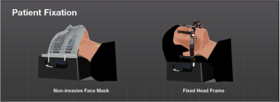
The traditional way of securing the patient’s head in stereotactic radiosurgery is by fastening an invasive head frame to the skull with sharp-tipped screws. Even though the head frame is effective for keeping patients immobile during treatment, some patients find head frame placement inconvenient and sometimes painful. Attaching the frame carries the risk of bleeding and infection, as well as requiring medication to be taken beforehand. The frame needs to remain in place for multiple hours, sometimes an entire day, until the treatment is completed.
With technologies like Image Guided Radiation Therapy (IGRT), “frameless” radiosurgery has become a popular alternative to invasive head frames for radiosurgery [2-4]. IGRT uses imaging technology during radiosurgery – X-ray, computed tomography (CT) – to both monitor the patient position and make any adjustments to the patient’s position and/or the radiation beams so that the teatment is precisely targeted to the tumor at all times.
Frameless radiosurgery is delivered using a non-invasive mask system. The mask is made from thermoplastic sheets that become soft when heated in water. Once formed on the patient, they cool in minutes and become hard again. The process is completely painless and typically requires no anesthesia. Prior to radiation, the mask is fitted to the patient and then fixated to the special treatment couch, keeping the patient immobile during the treatment. The process is similar to the way a frame-based treatment is delivered with the added benefit that no screws are attached to the skull, offering greater patient comfort.
There are different mask systems that use different techniques to ensure accuracy and monitor any patient movement during the radiation treatment.
Optical external surface tracking technology can be quite compatible with certain types of radiation treatments like breast cancer, and in fact most of the research studies surrounding accuracy in surface matching are based on breast cancer. However, it is a widely held belief that “submillimetric” treatment accuracy is critical when treating cancers in the brain.
In treating brain tumors, clinicians have the most demanding spatial tolerances of any site within the body. To accommodate the more limited precision of optical surface matching, clinicians may consider adding a ‘margin of error’ around the tumor that is being treated. However, the goal of any clinician is to minimize margins since expanding the area to be treated equates to knowingly treating healthy brain tissue. A ‘margin of error’ can significantly increase the total volume of the brain that is receiving a high dose of radiation. Patients must understand that this could potentially include vital structures that affect eyesight or hearing or balance. For example, adding a margin around the tumor border of just 2mm doubles the treatment volume of a brain metastasis that is only 15mm across.
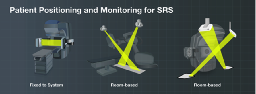
Patient Position Monitoring Technology
Patient position monitoring helps to maintain the accuracy of the procedure and ensure that the treatment dose is being delivered as prescribed by the cancer treatment team. Different delivery techniques offer different types of patient positioning and monitoring technologies.
When utilizing an approach with a head frame, doctors depend on various accessories and steps to align the tumor with the focal point of radiation. When a non-invasive mask is used, two low-dose X-ray images are captured from two different angles. They are compared and matched with simulated X-rays taken directly from the 3D CT data used for treatment planning. The robotic treatment table can fine-tune patient position with sub-millimeter movements. When you add imaging and micro-motion adjustments into the thermoplastic mask you can achieve the same level of accuracy as a frame based system without any of the discomfort of placing the frame. [7].
Ask your doctor about the different types of technologies and techniques to determine what the best procedure is for your brain metastases.
[1] Cosgrove, V. P. et al. Commissioning of a micro multi-leaf collimator and planning system for stereotactic radiosurgery. Radiother. Oncol. 50, 325–36 (1999).
[2] Gevaert, T. et al. Clinical Evaluation of a Robotic 6-Degree of Freedom Treatment Couch for Frameless Radiosurgery. Int. J. Radiat. Oncol. Biol. Phys. 83, 467–474 (2011).
[3] Cosgrove, V. P. et al. Commissioning of a micro multi-leaf collimator and planning system for stereotactic radiosurgery. Radiother. Oncol. 50, 325–36 (1999).
[4] Ackerly, T., Lancaster, C. M., Geso, M. & Roxby, K. J. Clinical accuracy of ExacTrac intracranial frameless stereotactic system. Med. Phys. 38, 5040–8 (2011).
[5] Pan, H. et al. Frameless, real-time, surface imaging-guided radiosurgery: clinical outcomes for brain metastases. Neurosurgery 71, 844–52 (2012).
[6] Kaul, D. et al. Dosimetric comparison of different treatment modalities for stereotactic radiosurgery of meningioma. Acta Neurochir. (Wien). (2014). doi:10.1007/s00701-014-2272-9
[7] Gevaert, T. et al. Setup Accuracy of the Novalis ExacTrac 6DOF System for Frameless Radiosurgery. Int. J. Radiat. Oncol. 82, 1627–1635 (2012).