Typically, a surgery that is navigated follows several steps:
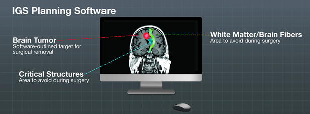
The patient’s diagnostic images like CT or MR are uploaded into the IGS system where the doctor can then create a plan for the surgery. This plan shows a full color, patient-specific 3D model of the brain tumor, the healthy tissue and critical brain structures near and around the brain tumor like the optic and cranial nerves and functional areas, responsible for hand and leg movement and the brain stem, for example.
This 3D model, which looks like an animation of the patient’s brain, is registered to the actual position of the patient on the operating table so that the surgeon can see or ‘track’ his instruments in relation to the patient’s real anatomy and orientate themselves on the 3D animation shown on the computer screen.
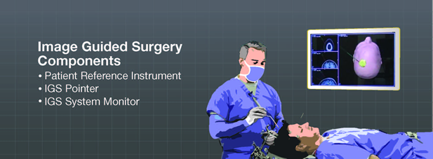
The patient can be tracked with different tracking technologies, which may include optical or electromagnetic. With the optical technology, the system requires special reflective markers on a reference instrument, which is placed close to or onto the patient’s head. These reflective markers are also located on the surgical instruments and are tracked by an infrared camera, which is connected to the system’s computer.
With all this information including size, location and orientation of the brain tumor, the surgeon can perform brain surgery while decreasing the risk to critical structures and healthy tissue.
Software
Now more than ever neurosurgeons are able to see an impressive amount of detail of the brain, not just structures like eyes and brain stem, but even fine and critical fibers that are the pathways of the brain. This visualization capability gives surgeons even more valuable intra-operative information.
Sophisticated software is used to allow the surgeon to see how different areas of the brain are connected and create a plan for treatment around critical areas. Software can also allow surgeons to more precisely outline a brain tumor as well as all the critical structures surrounding it. This helps better plan the surgical incision and visualize the best route to the tumor. This software is beneficial to surgeons because it helps take the guess out of the important areas of the brain that need to be avoided.
Using a Microscope during Image Guided Surgery
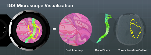
Image guided surgery offers a road map for surgeons to help plan out the shortest and safest way to access a brain tumor. Since neurosurgeons perform most of their procedures while looking through a microscope, microscope integration with image guided surgery is a very useful tool used during any brain procedure. It allows the neurosurgeon to switch between live images of the patient as well as images taken of the patient pre-surgery. Surgeons constantly need to know where they are during surgery. Before microscope integration, surgeons had to stop what they were doing and look at another navigation screen to find their location. Being able to view navigation information through the microscope while looking at the patient in real time saves the surgeon time during surgery, making it a valuable tool during surgery.