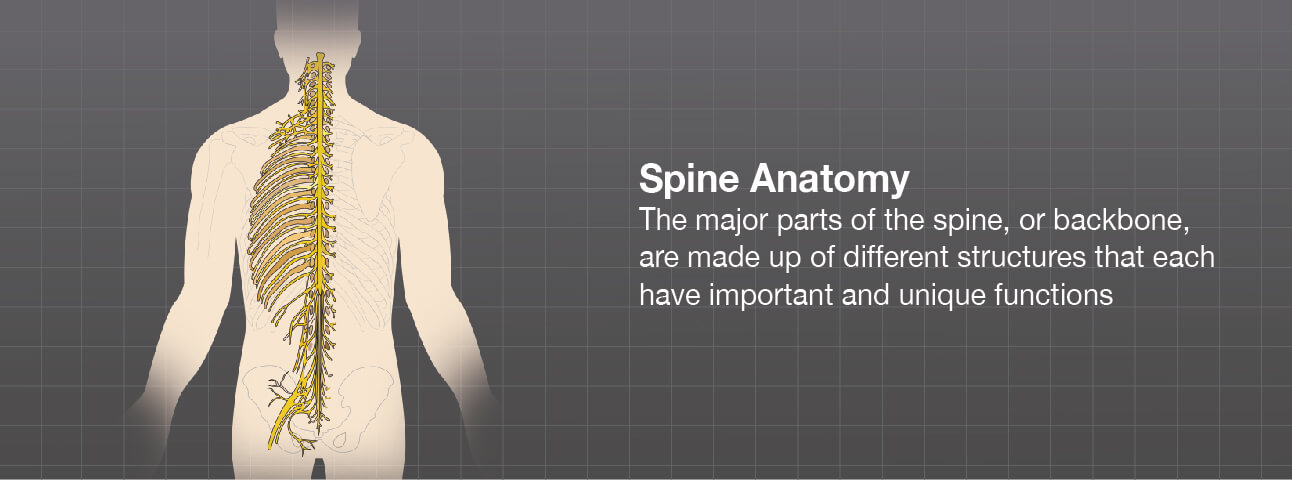
The back represents a very large part of the human body, starting at the base of the brain and extending from the neck and shoulders all the way down to the top of the buttocks. It’s breadth spans upper body from the shoulders down to the pelvis. It works as a single organ but is divided into three segments and different areas and structures with unique functions.
The spine is a physical support structure, holding up our head, shoulders and upper body. Our ability to stand up straight and flexibility to bend and twist come from the back. This complicated structure also protects the spinal cord, which is the all important communications system between the brain and the body.
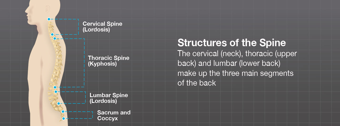
The cervical, thoracic and lumbar spine make up the three main segments of the spinal column, or backbone.
From the side, our three spinal column segments form natural curves. The ‘c-shaped’ neck and lumbar spine curves are called lordosis. The reverse ‘c-shape’ curve of the upper, or thoracic, spine is called kyphosis.
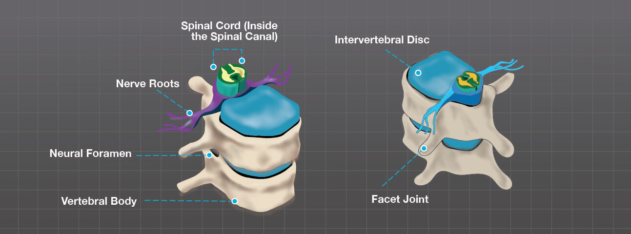
Each of the three major spine segments is made up of, and connected by, small rings of bones, called vertebrae. The vertebrae are stacked on top of each other and create a canal that houses the spinal cord. The bones serve to protect your spinal cord and, in turn, they are protected by a membrane that surrounds both the spinal cord and vertebrae.
Round, half-inch-thick discs separate the vertebrae from each other. These intervertebral discs move and expand and stabilize the spine, acting as shock absorbers. The discs allow us to move and yet maintain the spine’s strength at the same time.
Between the back of the vertebrae are facet joints. These small joints have a cartilage surface, like a hip or knee joint, and help your spine move and are important for spine rotation.
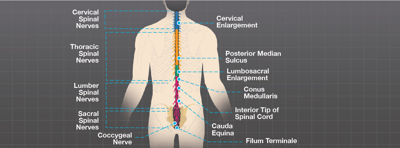
Spinal nerve cells, called neurons, carry messages to and from the spinal cord and between the brain and muscles, via a circuit of spinal nerves. Spinal nerves branch out from the spinal cord through openings in the vertebrae, called foramen.
The spinal cord ends around the first and second lumbar vertebrae in the lower back and continues as nerve roots. This bundle of nerve roots is called the cauda equina. They exit the spinal canal through openings in the vertebrae (foramen), just like other nerve roots. In the pelvis, some of the nerves group into the sciatic nerve, which extends down the leg.