Spine surgery requires high accuracy and precision technique. Oftentimes there is a very small incision so surgeons need to rely on intraoperative imaging to verify that they achieve the desired result.
When surgeons can view the bony anatomy and soft tissues in the spine, it helps them in their clinical decision making during the surgery and offers critical post-implant evaluation. Using images to review the surgical steps before closing the patient up and moving to recovery may increase safety and decrease re-operation rates.1
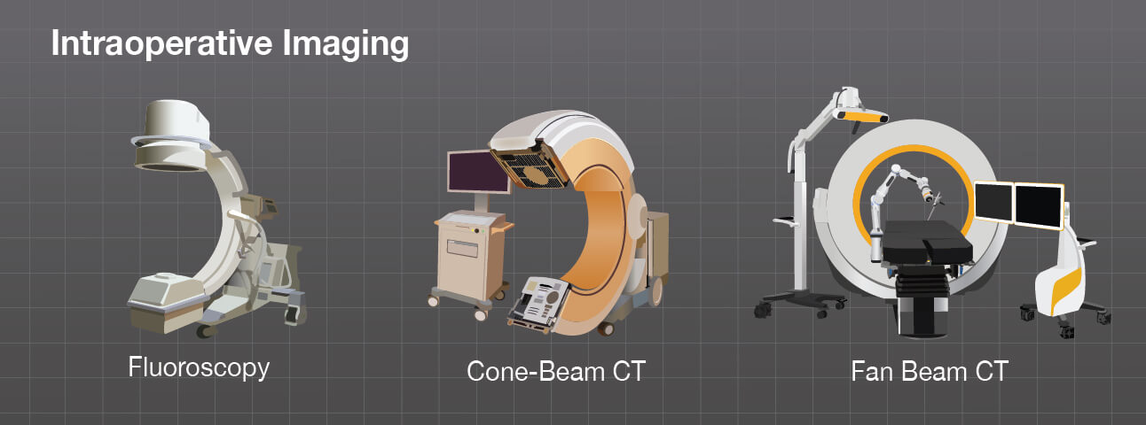
Traditionally, intraoperative verification of spine surgery steps has been done using fluoroscopy. While fluoroscopy offers reliable imaging, there is a concern about the level of radiation exposure to the patient, surgeon and operating room staff.
Newer intraoperative imaging technologies such as in-room 3D computed tomography (CT), combined with surgical navigation systems, greatly improve the way spinal surgeries are performed while drastically reducing X-Ray exposure for everyone in the operating room.2
1 Al-Khouja L, Shweikeh F, Pashman R, Johnson JP, Kim TT, Drazin D. Economics of image guidance and navigation in spine surgery. Surgical Neurology International. 2015;6(Suppl 10):S323-S326. doi:10.4103/2152-7806.159381.
2 Gebhard et al., Does computer assisted spine surgery reduce intraoperative radiation doses?
Spine (Phila Pa1976). 2006 Aug 1;31(17).