Surgical navigation systems are used to both plan and execute spine procedures. The technology has advanced a great deal over the last decade, enabling surgeons to assess the surgical case, pre-plan the procedure and achieve reproducible outcomes.1
Surgical navigation drives access to real-time imaging and enriched patient data that surgeons can use to make timely and informed decisions, as well as obtain pre-closure verification of results.
Pre-Planning
The surgical team uses the software before the surgery to enrich images that are uploaded to the system. Doctors are able to review CT scans to identify bone structures and plan surgical steps. If magnetic resonance imaging (MRI) scans were taken, doctors may upload and use those images to define tissue, outline critical structures and identify a tumor or lesion. The system even allows surgeons to fuse CT and MRI images together to get an even more complete picture of the anatomy and to plan and execute the procedure.
Intraoperative
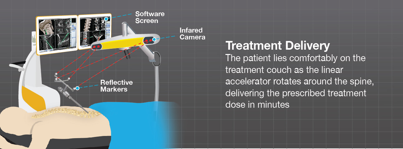
Once the surgery is planned, the software is used to create a computerized model of the patient’s spine, which resembles a 3D anatomical animation. During the procedure, the navigation system displays patient data collected in real time during surgery, helping to guide the surgeon through important surgical steps. Surgeons are able to view images of the spine, discs and nerves during surgery and confirm placement with submillimetric accuracy.2
By ‘tracking’ patient anatomy, surgical instruments and artificial components like implants in relation to patient anatomy, image guided surgery systems support doctors in measuring and completing all of the planned steps to restore the spine.
1 15 Things to Know about Robotic Spine Surgery, Becker’s Spine Review, March, 2017.
2 McLaughlin, M., Image Guided Spine Surgery, Spine Universe. Mar. 4, 2016.