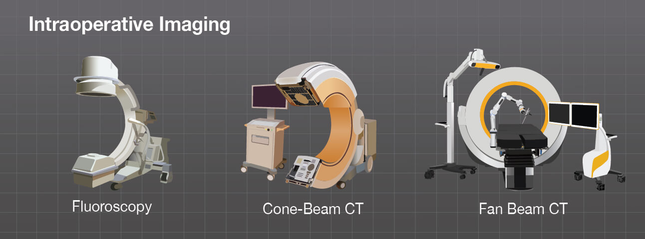
Intraoperative Imaging
Most spine surgeries today are done using minimally invasive techniques to spare muscle and healthy tissues. To do this as effectively as possible, some form of intraoperative imaging is typically used to verify surgical accuracy. The intraoperative images help make sure that a spinal implant is placed in the desired place or that a tumor is dissected to the desired outcome.
The traditional way of verifying spine surgeries is with intraoperative fluoroscopy. While fluoroscopy offers reliable imaging, there is a concern about the level of radiation exposure to the patient, surgeon and operating room staff.
The evolution of the in-room mobile computed tomography (CT) scanner combined with the advancements in imaging and computer-based navigation have considerably improved accuracy over that of traditional fluoroscopy-guided techniques, particularly during pedicle screw positioning. In one study, use of a mobile CT scanner reduced the rate of screw repositioning, which enhanced patient safety and diminished radiation exposure for patients.1
While providing excellent imaging resolution and navigation to guide an operation, the mobile CT scanner also permits the surgeon to obtain immediate CT images at the completion of surgery. This allows for immediate intraoperative intervention if necessary before surgical closure.2
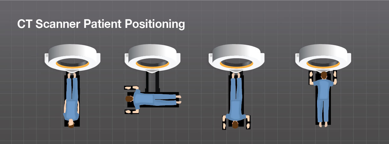
Before an image is taken with a mobile CT scanner, most staff moves out of the area, or even the room, which limits radiation exposure. Once the image is obtained, it is automatically registered to the patient scans in the navigation software, helping the doctor verify that the implant is placed correctly or the tumor is resected as desired. The use of high quality images offers critical knowledge and increases surgeon confidence, playing an important role in spine surgery and may help reduce the rate of re-operation in some cases.
There are systems that use slightly different ways to orient the patient with the surgical navigation system. These optical surface matching techniques are suitable in many cases, especially minimally invasive percutaneous surgeries, but have their drawbacks including skin movement, incision and operation site size.
1 Journal of Neurosurgery: Spine.
2 Doniel Drazin, M.D., Terrence T. Kim, M.D., David W. Polly Jr, M.D., and J. Patrick Johnson, M.D., Introduction: Intraoperative spinal imaging and navigation, Neurosurgical Focus, Mar 2014 / Vol. 36 / No. 3.