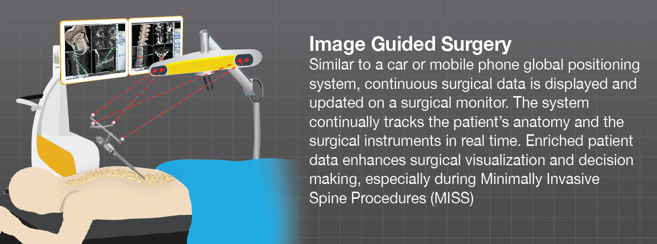
Image guided surgery (IGS), also known as surgical navigation or computer-assisted navigation (CAN) is a method that neurosurgery and orthopedic surgeons may use during spine procedures.
Similar to a car or mobile Global Positioning System (GPS), image guidance systems continuously track points of the anatomy and display them on a computer monitor in the operating room before, during and after surgery, helping to guide the surgeon through important milestones of the procedure. Surgical navigation provides your doctor with additional information and measurements and tracks the surgical instruments being used for the procedure.
For many years, intraoperative navigation was used primarily for pedicle screw placement to help stabilize the spine. Now, surgeons rely on image guidance for many other procedures, including surgical resection of intradural and spinal column tumors, cases of infection, revision procedures on arthrodesed spines, and deformity cases with distorted anatomy. Beyond visualization during surgery, navigation platforms may mitigate much of the harmful radiation exposure in minimally invasive surgery (MIS) to which the patient, surgeon, and ancillary operating room (OR) staff are subjected.1
In 2011, more than 740,000 people underwent spinal surgery in the United States, with spinal fusion, discectomy, spinal device implantation and spinal decompression the most common of those procedures.2
Surgeons trust advanced technologies like image guidance because they offer high definition visualization of intraoperative patient data. This system of checks and balances throughout a procedure is critical because spinal procedures are often performed through small incisions designed to be minimally invasive, limit muscle damage and protect structures near the operative site. Studies show that image guidance platforms have been shown to dramatically improve a surgeon’s manual dexterity allowing for greater control and maneuverability through a less invasive working portal, while dampening a surgeon’s physiological tremor.1
There are times when the patient data, or images, do not offer high definition visuals of the area of the spine where a procedure needs to be performed. In these cases, as well as those where minimally invasive techniques are used, surgical navigation may potentially allow for a more accurate and secure implant placement compared to procedures without intraoperative planning.3 Using the navigation software, surgeons are able to build a clear reconstruction of the patient’s CT and/or MRI images, then plan and execute the desired surgical path for accurate placement of the implant. Surgeons can also react and verify intra-operatively and, if necessary, correct performed treatment steps during surgery. Accurate placement and stability of spinal implants is critical to overall back function and can potentially help the implant to last longer.
In other spine surgeries, like tumor removal, image guidance systems offer powerful software that helps to outline, or contour, the tumor as well as critical surrounding structures, like nerves and the spinal column. Once planned, surgeons are able to track their movements on the system’s computer monitor and follow the mapped out route to the tumor and resect, or remove, as much, or all, of the tumor as planned.
The application of image guidance navigation techniques to address simple or complex pathologies has translated into better outcomes and faster recovery in all areas of the spine.3 Multiple studies1 demonstrate why doctors and surgical staff rely on computer-assisted navigation.
The Concerns with Intraoperative Fluoroscopic Imaging
Traditionally, doctors doing spine surgeries used a type of medical imaging called intraoperative fluoroscopy. Fluoroscopy offers a continuous X-ray image—like an X-ray movie—which is possible because the X-ray beam is passed through the patient’s body. The fluoroscopy images are used to help guide and localize the spine instruments for fusion procedures.
However, the X-rays used can be considered hazardous radiation exposure that affects patients, surgeons and O.R. staff and are much higher for spine procedures than for other subspecialties in orthopedic and neurosurgery.1
During minimally invasive spine surgeries (MISS), surgeons rely even more on intraoperative fluoroscopy to help guide their instruments.
The advent of surgical navigation and newer intraoperative imaging technologies like cone beam CT and CT have helped decrease the exposure and effects of fluoroscopic radiation.
Visit our What is Intraoperative Imaging tab in this same section to explore additional spinal surgery technologies.
Talk to your neurosurgeon or orthopedic surgeon to find out if image guided spine surgery is an option for you.
1 Samuel C. Overley, Samuel K. Cho, Ankit I. Mehta, Paul M. Arnold; Navigation and Robotics in Spinal Surgery: Where Are We Now?, Neurosurgery, Volume 80, Issue 3S, 1 March 2017, Pages S86–S99.
2 http://www.boneandjointburden.org/2014-report/iie0/spine-procedures
3 Doniel Drazin, M.D., Terrence T. Kim, M.D., David W. Polly Jr, M.D., and J. Patrick Johnson, M.D., Introduction: Intraoperative spinal imaging and navigation, Neurosurgical Focus, Mar 2014 / Vol. 36 / No. 3.